jueves, 28 de febrero de 2019

Association between OSA and cardiovascular diseases in women

Association between OSA and cardiovascular diseases in women

News-Medical

Association between OSA and cardiovascular diseases in women

In the current issue of Cardiovascular Innovations and Applications (Special Issue on Women's Cardiovascular Health, Volume 3, Number 4, 2019, Guest Editor Gladys P. Velarde) pp. 421-434(14); DOI https://doi.org/10.15212/CVIA.2017.0064, Nimeh Najjar, MD, Peter Staiano, MD and Mariam Louis, MD from the University of Florida, Department of Medicine, Jacksonville, FL, USA consider obstructive sleep apnea and cardiovascular disease in women.
The authors review important sex differences in the association of Obstructive Sleep Apnea (OSA) and CVD with some data suggesting women may have different susceptibility to the effects of OSA. The authors stress that although OSA is a common condition, it remains grossly underdiagnosed in women.
Obstructive Sleep Apnea (OSA) is a common chronic disorder that affects 5-10% of the US population with a higher prevalence in men compared to women by 2:1 in population studies. By contrast, in patients with cardiovascular disease, its prevalence can exceed 50% depending on the specific disorder surveyed. Although sex differences have been well described for cardiovascular risk factors; existing data regarding the impact of sex on the relationship between OSA and cardiovascular outcomes is controversial. Similarly, while there is strong evidence for increased prevalence of cardiovascular conditions, such as systemic hypertension, atrial fibrillation, as well as heart failure amongst patients suffering from OSA; conflicting evidence exists regarding the incidence and bidirectional relationship between them as well as the impact of treatment of OSA on cardiovascular outcomes. In this paper, the authors review the associations between OSA and cardiovascular diseases in women. The data on sex differences is limited, due to a number of reasons, including, but not limited to late presentation of OSA in women, difficulties in diagnosing both OSA and cardiovascular diseases in women and still suboptimal inclusion of adequate number of women in clinical trials. More studies are needed to better delineate sex differences in the clinical presentation as well as the pathophysiology of the associations between OSA and cardiovascular diseases so that we can provide patients with more personalized care.
This article forms part of a special issue on Women's Cardiovascular Health, guest edited by Gladys P. Velarde. Recent decades have witnessed great progress in the treatment of cardiovascular disease (CVD). Due to improved therapies, preventive strategies and increased public awareness, CVD (stroke, heart failure, ischemic heart disease, peripheral arterial disease and congenital heart disease) mortality has been on the decline over this span of time for both genders. Unfortunately, the decline has been less prominent for women, especially women of color. Once viewed as a man's disease, CVD remains the leading cause of mortality for women in the United States and is responsible for a third of all deaths of women worldwide and half of all deaths of women over 50 years of age in developing countries. In the United States, CVD far outpaces all other causes of death, including all forms of cancer combined. The statistics are sobering with about one female death in the United States every 80 seconds from CVD. That represents close to 400,000 deaths per year according to the more recent statistics. Of these, more than one quarter of a million women will die this year from ischemic heart disease (IHD) which includes obstructive and non-obstructive coronary disease, and about 64% of women who die suddenly of IHD have no prior symptoms. Despite a significant number of females with known CVD and increased awareness among women of heart disease as their major health threat, a substantial proportion of women (46% as per the most recent American Heart Association survey) remain unaware of their cardiovascular risk and continue to fail to recognize its significance.
This lack of awareness is more profound (over 60% unaware) among women in higher-risk groups, racial and ethnic minorities, and has changed little in decades.
Poorly understood sex/gender differences in pathobiologic mechanisms, clinical presentation, management and application of diagnostic and therapeutic and preventive strategies have contributed to this gap. A critically important factor has been the underrepresentation of women in CVD research to date. In fact, only one-third of CVD clinical trials report sex-specific results despite The Food and Drug Administration regulations requiring sex stratification data, as well as the National Institute of Health recommendations of increased inclusion of women in clinical trials. This makes it difficult for researchers and clinicians to draw accurate conclusions about sex differences in mechanisms of disease, accuracy of specific diagnostic modalities and risks or benefits of a particular drug or device for the treatment of women with CVD. Furthermore, physicians and other healthcare providers continue to underestimate women's cardiovascular risk, in part because of utilization of traditional approaches which can lead to over-testing or inappropriate risk assessment without accurate differentiating who is truly at risk and inadequate use of preventive therapies for women.
The goal of this special edition Cardiovascular Innovations and Applications is to shed some light on specific topics that dominate the spectrum of CVD in women.

Extra weekend sleep cannot repair the damage from sleep-deprived work week

Extra weekend sleep cannot repair the damage from sleep-deprived work week

News-Medical

Extra weekend sleep cannot repair the damage from sleep-deprived work week

On some measures, a yo-yo sleep schedule can be worse
Think sleeping in on the weekend can repair the damage from a week of sleepless nights?
Not so, according to University of Colorado Boulder research published today in Current Biology.
In fact, on some health measures, trying to play catch-up for a few days and then returning to poor sleep habits makes things worse.
"Our findings suggest that the common behavior of burning the candle during the week and trying to make up for it on the weekend is not an effective health strategy," said senior author Kenneth Wright, director of the Sleep and Chronobiology Lab.
Previous research has shown that insufficient sleep can boost risk of obesity and diabetes, in part by boosting the urge to snack at night and decreasing insulin sensitivity - or the ability to regulate blood sugar. Some adverse health impacts kick in after just one night of lost sleep, recent CU Boulder research has shown.
Sleeping in on the weekend can help the body recover mildly during those two days, studies suggest. But the effects don't last.
Wright and lead author Chris Depner, an assistant research professor of Integrative Physiology, wanted to determine what happens when people cycle back and forth between a sleep-deprived work week and a few days of catch-up.
They enlisted 36 healthy adults age 18 to 39 to stay for two weeks in a laboratory, where their food intake, light exposure and sleep were monitored.
After baseline testing, the volunteers were divided into groups. One was allowed plenty of time to sleep--9 hours each night for 9 nights. The second was allowed 5 hours per night over that same period. The third slept no more than 5 hours nightly for 5 days followed by a weekend when they could sleep as much as they liked before returning to 2 days of restricted sleep.
Both sleep-restricted groups snacked more at night, gained weight and saw declines in insulin sensitivity during the study period. While those in the weekend recovery group saw mild improvements (including reduced nighttime snacking) during the weekend, those benefits went away when the sleep-restricted work week resumed.
"In the end, we didn't see any benefit in any metabolic outcome in the people who got to sleep in on the weekend," said Depner.
On some measures, the weekend recovery group showed worse outcomes. For instance, in the group which had their sleep restricted the whole time, whole body insulin sensitivity declined by 13 percent. In the weekend recovery group it worsened by 9 to 27 percent, with sensitivity in the muscles and liver scoring worse than the other groups.
"It could be that the yo-yoing back and forth - changing the time we eat, changing our circadian clock and then going back to insufficient sleep is uniquely disruptive," said Wright.
Even when given the chance, people found it difficult to recover lost sleep. While they gained some ground Friday and Saturday, their body clocks shifted later Sunday night making it hard to fall asleep even though they had to get up early Monday.
In the end, the recovery group got just 66 minutes more sleep on average. Men made up more lost sleep than women.
Wright says it's possible that weekend recovery sleep could be an effective health countermeasure for people who get too little sleep a night or two per week. They hope to explore this in future studies.
For now, the takeaway is this: Consistency matters.
"This study demonstrates the importance of getting sufficient sleep on a regular schedule," said Michael Twery, Ph.D., director of the National Center on Sleep Disorders Research (NCSDR) at the National Heart, Lung, and Blood Institute, part of the National Institutes of Health. "Frequently changing sleep schedules is a form of stress associated with metabolic abnormalities."
So try to get 7 hours of sleep as many nights as possible.

Stress controllability mitigates the negative effects

Stress controllability mitigates the negative effects

News-Medical

Stress controllability mitigates the negative effects

In individuals, stress exposure in adolescence increases vulnerability and risk of developing psychopathologies in adulthood, such as drug addiction, mood, anxiety, addiction to gambling, attention deficit hyperactivity disorder, etc. Researchers at the Universitat Autonoma de Barcelona observed in animal models that the ability to control the source of stress diminishes its effects and could reduce the risk of later developing mental disorders. The research appears today in the journal Scientific Reports.
Researchers from the Institute of Neuroscience of the Universitat Autònoma de Barcelona (INc), led by Roser Nadal and Antonio Armario, conducted a study on the factors which reduce the effects of stress. The research, appearing today in the journal Scientific Reports,used three groups of male rats to measure these effects. One group underwent several sessions of stress during their adolescence, which they could control (by stopping or preventing) by acting in a certain manner. A second group received the same amount of stress sessions as the first group, but their behavior had no effect (uncontrollable stress). A third group acted as a control group and underwent no stress.
During the exposure to stress, researchers quantified the intensity of their reaction by measuring the endocrine response through the activity of the hypothalamic-pituitary-adrenal axis (HPA axis). In the adult stage, several experiments were conducted to measure different cognitive variables and the expression of dopamine type 2 receptors in the dorsal striatum, an area of the brain relevant to the behaviours measured. Part of these data forms part of the PhD thesis of INc researcher Maria Sanchís Ollé, first author of the paper.
The results indicated that HPA activation induced by controllable and uncontrollable stress was the same in the first exposure to stress. However, with repeated exposures the controllable stress group demonstrated an attenuated HPA response. In their adult stage, the animals exposed to uncontrollable stress in adolescence developed an increase in motor impulsivity and a decrease in cognitive flexibility, effects which were not made evident in those animals exposed to controllable stress. Other aspects (attention and cognitive impulsivity) were not observed to have been affected by stress. At the same time, the behavioral effects of uncontrollable stress were associated with an increase of the number of dopamine type 2 receptors in the dorsal striatum (but not in other sub-divisions), a structure involved in impulsivity and cognitive inflexibility.
"Despite the fact that being exposed to situations of stress has short and long-term negative effects on behavior and physiology, there are several factors which could mitigate its impact. We have observed that one of these factors is the possibility of having control over the source of stress", affirms Roser Nadal.
The study has several preventive implications and points to the fact that strategies aimed at increasing the perception of stress controllability during adolescence could mitigate the negative effects of stressful experiences in the adult age and reduce vulnerability to certain psychopathologies.

Optic Disc Swelling: Overview

Optic Disc Swelling: Overview

News-Medical

Optic Disc Swelling: Overview

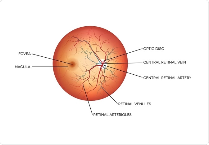
The optic disc is the round spot on the retina formed by the passage of the axons of the retinal ganglion cells, which transfer signals from the photoreceptors of the eye to the optic nerve, allowing us to see.
Diagram of the eye, showing the optic diskTefi | Shutterstock
The optic disc, also known as the optic nerve head, forms a slightly raised spot on the nasal side of the retina. Here, there are no photoreceptors, hence it is known as the blind spot of the eye. The optic disc has a slight cup-shaped depression, called the physiologic cup. The central retinal artery and vein also pass through the retina at this point. Swelling of the optic disc limits vision and can have a significant effect on quality of life.

Causes of optic disc swelling

Swelling of the optic disc can occur in reaction to a variety of insults. The most common causes of swelling include:
  • Non-arteritic anterior ischemic optic neuropathy (NA-AION): This condition affects over one third of patients and is characterized by the loss of blood to the optic nerve, resulting in ischemia and cell death.
  • Optic neuritis: This condition accounts for just less than one third of pateints, and is caused by inflammation around the optic nerve.
  • Intracranial hypertension: Affecting one in six patients, intracranial hypertension occurs when fluid builds up in the brain, putting pressure on the eye.
  • Other causes include diseases such as diabetes, where optic disc swelling occurs as a secondary disease.

Unilateral or bilateral swelling

Swelling of the optic disc may be confined to one side (unilateral) or occur on both sides (bilateral) of the retina. Unilateral swelling is observed in the following disorders, among others:
  • Demyelinating optic neuritis
  • NA-AION
  • Compressive optic neuropathy
  • Occlusion of the retinal vein
  • Diabetic papillopathy
Bilateral swelling of the optic disc may be due to causes such as:
  • Papilledema
  • Infiltrative neuropathy of the optic nerve
  • Toxic optic neuropathy
  • Malignant hypertension

Symptoms

Swelling of the optic disc presents with symptoms based on the underlying causative condition. In the majority, therefore, patients complain of a sudden diminution of visual acuity and visual field. Visual field defects in NA-AION are mostly inferonasal in location. Altitudinal field defects, affecting only half of the vertical field of vision, are more common in optic neuritis, but scotomas of different types or an enlargement of the blind spot are also often present.
Pain on eye movement is seen more frequently in optic neuritis, but is typically absent in ischemic neuropathy. In compressive forms of optic neuropathy, various intracranial tumors should be ruled out. A relative afferent pupillary defect is characteristic, but pain is usually absent.

Diagnosis and Treatment

The following signs indicate the presence of non-papilledema optic disc swelling:
  • The cupping of the disc is lost
  • The margins of the disc are blurred
  • No peripapillary hemorrhages are seen
  • No infarcts, exudates or edema of the nerve fiber layer
Diagnosis depends on a careful history of symptoms related to intracranial hypertension, other systemic conditions and visual symptoms related to the current episode.
Tests include dilated fundoscopy and imaging findings obtained with fluorescein angiography and optical coherence tomography (OCT). Treatment of optic disc swelling is based on the cause of the condition.

Further Reading

Last Updated: Feb 28, 2019

Recognizing the Signs of Major Depressive Disorder (MDD)

Recognizing the Signs of Major Depressive Disorder (MDD)

News-Medical

Recognizing the Signs of Major Depressive Disorder (MDD)

Major Depressive Disorder (MDD), also known as clinical depression, is a psychiatric condition that affects more than 15.7 million adults and approximately 3 million adolescents (aged 13 to 17 years) in the USA each year.
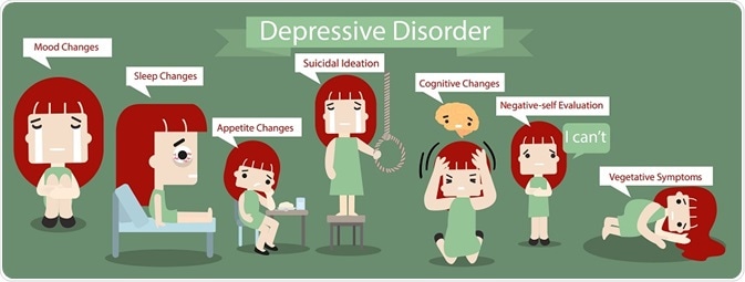
illustration of the main symptoms of major depressive disorder24cdesign | Shutterstock
The World Health Organization estimates that more than 300 million are affected by depression globally. Depression may be caused by adverse environmental experiences (e.g. loss of a loved one), biochemical imbalances (e.g. low serotonin levels), or neurological responses to stressful stimuli (e.g. overactivity of the HPA axis).
While MDD can present in multiple forms, signs and symptoms can vary between individuals and may be difficult to differentiate from other forms of depression.

Diagnostic criteria of MDD

According to the 5th edition of the Diagnostic and Statistical Manual of Mental Disorders (DSM-5), a person diagnosed with MDD meets at least five of the following conditions for a period of at least two weeks:
  • Depressed mood most of the day
  • Loss of interest or pleasure (anhedonia)
  • Significant weight loss or weight gain
  • Insomnia or hypersomnia almost everyday
  • Observable psychomotor agitation (feelings of restlessness)
  • Fatigue or loss of energy
  • Feelings of worthlessness or excessive guilt
  • Reduced capacity to concentrate or think
  • Recurrent thoughts of death or suicidal tendencies
It is essential to note that for an individual to be diagnosed with MDD, they must experience one of the first two symptoms plus at least four of the other symptoms. Clinical depression significantly affects the daily behavior and personality of an individual and this is usually noticed by collegues, family members and people close to the patient.

Functional changes

People with clinical depression experience significant functional changes in most of their social and personal commitments. Functionality would refer to any form of activity that enables an individual to take care of themselves and establish independence in accomplishing simple day-to-day activities. A person with clinical depression would have markedly lower functional abilities, especially in terms of personal care.
Studies have shown that individuals with MDD are less likely to care for personal grooming and hygiene. They would also most likely lack the motivation and capacity to attend school or report to work. These functional changes are significantly observable and would facilitate grave limitations in terms of the individual’s capability to follow their usual day-to-day routine.

Suicidal tendencies

Recurrent thoughts of death or suicidal ideation is an important, but not necessary sign for the diagnosis of clinical depression. Affected persons may attempt suicide, and, physicians should treat anyone with suicidal thoughts as a medical emergency that requires immediate attention and support.
In clinical practice, physicians and psychologists assess for the  escalation of  depressive and suicidal thoughts by asking a person how they are likely to act on their thoughts. It is important to distinguish between passive thoughts of “wanting to die” from the more serious immediate risk associated with having plans for committing suicide.

Anhedonia

Another significant marker of major depressive disorder is significant loss of interest or pleasure. Medically termed anhedonia, the condition is marked by low excitement and happiness in association with activities that would typically bring joy and satisfaction to the patient. Thus, a person experiencing a depressive state may have reduced capacity to seek pleasure or enjoyment on various social and environmental encounters. The DSM-5 notes that either anhedonia or depressed mood are required for  a diagnosis of MDD.

Further Reading

Last Updated: Feb 28, 2019

Preventing Ocular Hypertension

Preventing Ocular Hypertension

News-Medical

Preventing Ocular Hypertension

Ocular hypertension is defined as a higher than normal intraocular pressure (eye pressure) that significantly increases the risk of developing glaucoma.
Image of the human eyeRocketclips, Inc. | Shutterstock

Epidemiology of ocular hypertension

The incidence of ocular hypertension varies among by race and ethnicity, and also increases with age. According to one epidemiological study, African-Caribbean persons are more at risk for ocular hypertension; this group has the highest prevalence of disease globally (12.6%).
Among White persons enrolled in the Framingham Eye Study, prevalence was 6.2% in persons aged less than 65 years and 8.7% in persons aged older than 75 years. In South India, the prevalence is 1.1% in persons older than 40 years.

Why is it important to detect ocular hypertension early?

Although ocular hypertension is mostly asymptomatic, chronically high eye pressure can trigger glaucomatous optic neuropathy (disc changes and visual field defects). According to the Ocular Hypertension Treatment Study (OHTS), about 2% of people with untreated ocular hypertension develop glaucoma in one year, and 9.5% develop glaucoma in 5 years. After 13 years, that rises to 22%.
An increased intraocular pressure is the most frequent risk factor for glaucoma (a serious eye disorder characterized by optic nerve damage and vision loss); however, glaucoma can also develop in the absence of ocular hypertension (low-tension or normal-tension glaucoma). Several factors that are associated with ocular hypertension can further increase the risk of glaucoma onset.
Central corneal thickness is one such risk factor. The accuracy of the eye pressure measurement significantly depends on the biochemical properties of the cornea. In case of thicker cornea, applanation tonometry may provide an overestimated value of the eye pressure; whereas, in case of thinner cornea, the value may be underestimated.
Every 40 µm decrease in central corneal thickness increases the risk of glaucoma development by 81%. Therefore, while evaluating glaucoma risk factors, it is important to keep in mind that people with thinner corneas and ocular hypertension are more susceptible to glaucoma than those with thicker corneas and ocular hypertension.
Another risk factor is cup-to-disc ratio, which is used to assess the progression of glaucoma. The optic cup is the cup-like central portion of the optic disc. In glaucoma, higher intraocular pressure causes additional cupping of the disc, which is a pathological eye condition.
The cup-to-disc ratio measures the difference between the diameter of cup-like portion of the optic disc and the total diameter of the optic disc. An increase in horizontal and vertical cup-to-disc ratio can significantly increase the risk of glaucoma progression.

Is ocular hypertension preventable?

Steps can be taken to prevent ocular hypertension becoming glaucoma, and this is particularly important in patients with a family history of the condition. Treatment consists of topical eye drops which reduce the eye pressure and thus, glaucoma risk by 50%.
Regular monitoring of the eye pressure is also important. Moreover, patients with ocular hypertension must undergo annual follow-ups with visual field testing and optic nerve imaging.    
Given that medications can be expensive and side effects and poor adherence are possible, it is important to categorize patients with ocular hypertension as low, moderate, or high risk before initiating treatment.
Patients with eye pressure of > 30 mm Hg are particularly at high risk. Similarly, patients with eye pressure of > 26 mmHg and central corneal thickness of < 555 microns are at high risk. These individuals definitely require medications to reduce the eye pressure by at least 20%.
An eye pressure of 24 – 29 mm Hg without retinal nerve damage or an eye pressure of 22 – 25 mm Hg with central corneal thickness < 555 microns is considered moderate risk. Annual follow-ups are essential for patients who are at moderate risk of glaucoma. Treatment for this patient cohort generally starts only after the diagnosis of early glaucoma-related changes.  
Patients with eye pressure of 22 – 23 mm Hg and central corneal thickness of > 588 microns are at low risk and rarely develop glaucoma. Thus, treatment is not recommended for these patients. However, they should undergo a follow-up every 2 years.

Further Reading

Last Updated: Feb 28, 2019

MD&M West 2019: Medical Devices, 3D Printing and Staying Compliant

MD&M West 2019: Medical Devices, 3D Printing and Staying Compliant

News-Medical

MD&M West 2019: Medical Devices, 3D Printing and Staying Compliant

Between February 5 - 7 2019, the Anaheim Convention Center in Southern California hosted the annual MD&M West medical manufacturing conference.
This year, MD&M organizers chose to highlight the latest advancements in medical device technology and innovative marketing strategies that can be used to target specific consumer audiences. The conference also featured updates to recent regulations for newly-developed medical devices in both the United States and Europe.


Innovative 3D Printing Solutions

The incorporation of three-dimensional (3D) printing, also referred to as additive manufacturing, has continued to intensify across a wide variety of industries, as companies around the world become increasingly aware of how cost-effective this technology is.
Within the medical field, researchers are starting to use 3D printing to create tissues and organoids as potential alternatives to human organ transplants. Furthermore, 3D printing has also improved medical teaching curricula by creating patient-specific organ replicas for surgeons to practice on before performing complicated procedures.
This year’s MD&M West event explored these types of 3D printing innovations by discussing how various 3D printing healthcare applications have transitioned from a prototyping phase to mainstream production. Speakers like Grant Shirley from Stratasysargued the importance of maintaining rigorous quality assurance testing and inspection processes of all 3D printed materials, particularly those which exhibit complex geometries and are composed of new and different materials.

Supporting Product Development

Example of bioprintingstockddvideo | Shutterstock
As technology companies continue to make their mark within the clinical setting, it is imperative that product designers and manufacturers maintain a close relationship with their end users who, in this case, are the clinicians and patients that will be benefitting from these products.
Many technology companies that have experience with developing medical devices agree that, in addition to prototyping their devices through numerous usability tests, active conversation with other companies, as well as clinicians and patients, was pivotal to their success.
The opening plenary to MD&M West’s Product Development & User-Centered Design track provided a unique insight into the entrepreneurs attending the conference on the importance of collaboration in this field.
Anne Wellington, who is currently a managing director at Cedars-Sinai, informed the audience on how young companies can collaborate with their prospective end users of the health industry to develop products that precisely meet their consumers’ needs.
Phillip Remedios of BlackHagen Design also supported this discussion in a separate seminar by examining the methodology needed to achieve innovative product design.

Staying Up to Date with the EU and the FDA

Engineers designing a medical deviceStock-Asso | Shutterstock
In 2018 alone, over 50 different types of medical devices were approved for use by the United States Food & Drug Administration (FDA). These novel medical devices range from ophthalmic surgical lasers, heart pumps, implantable lenses, glucose monitoring systems and much more.
At MD&M 2019, Dr. Daniel Shultz discussed the FDA’s current strategies and areas of concerns regarding medical device technology, and how the priorities of this federal entity have the potential to affect the manufacturing companies that produce these products.
In addition, Dr. Michael Drues discussed the various programs that have been incorporated into the FDA’s Breakthrough Designation Program (BDP). In an effort to improve patient access to novel technologies, Dr. Drues discussed how manufacturers can apply for BDP designation and tips on how to ensure that their applications are not rejected or delayed.
Although the regulations set by federal entities within the United States and the European Union are primarily interested in the safety and well-being of patients, medical device manufacturers may feel restricted by these laws. For example, the Restriction of Hazardous Substances (RoHS) and Registration Evaluation and Authorization [and restriction] of Chemicals (REACh) acts of the European Union have continued to increase restrictions on medical devices that have been miniaturized or those that are composed of more exotic materials.
To overcome these legislative challenges, Thomas Bowles of FUTEK Advanced Sensory Technology, Inc. discussed ways in which product designers can meet the demands of these restrictions while also allowing their products to progress to market.

Conclusion

Overall, the 2019 MD&M West conference provided attendees with numerous different sessions that covered a wide variety of relevant topics on product trends, new materials, artificial intelligence, robotics and much more.
With respected speakers from highly successful companies, such as Edwards Lifesciences, Galen Robotics and Merck, as well as health professionals from well-renowned hospitals like Mayo Clinic, this year’s MD&M West provided attendees with an intimate view on the future of medical technology.